1. The birth of medical X-ray machine: a hundred years from laboratory to operating room
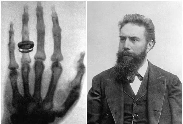
On November 8, 1895, German physicist Wilhelm Rontgen discovered a mysterious ray that could penetrate books, wood, and even human bodies while experimenting with a cathode-ray tube in his laboratory. Due to its unknown nature, Rontgen named this ray “X-rays.” He used X-rays to take a photograph of his wife’s hand bone, which clearly showed the outline of the bone and the ring. This photograph became a landmark in medical history.

2. The “growth history” of X-ray machines in surgical applications: from post-confirmation to real-time guidance
- Early stage (early 20th century to 1980s): X-ray was only used for preoperative or postoperative radiography to assist in judging the reduction of fracture or the position of foreign bodies. It could not be involved in real time during surgery, and doctors had to rely on experience to operate, with a high error rate.
- Digital transformation (1990s-2010): CR (computer radiology imaging) and DR (digital radiology imaging) technologies are popularized, X-ray machines can realize digital imaging, and images can be quickly obtained through flat panel detectors during surgery, but the equipment still needs to be fixed and installed, and the mobility is limited.
- Portable Breakthrough (After 2010): As medical needs evolved towards “precision and minimally invasive” procedures, real-time intraoperative imaging guidance became essential. Portable X-ray machines emerged to meet this demand, compressing the device size to a portable level. Combined with wireless transmission technology, they allow doctors to access high-definition images at any time during surgery, completely transforming the traditional “blind operation” model.
3. Technological innovation of portable X-ray machine: Bojin is at the forefront
- Miniaturized design: using integrated ball tube and detector, the weight is as low as 9 pounds (about 4.1kg), some models support handheld operation, with wheels can flexibly shuttle in the operating room, ward.
- Low radiation and high clarity: Through micro-focus ball tube (0.4mm focus) and short exposure technology (minimum 0.02 seconds), the resolution of 50LP/cm can be achieved while reducing the radiation dose by more than 80%, and the fine structures at the millimeter level (such as fracture lines in children, foreign bodies in soft tissues) can be clearly displayed.
- Intelligent interconnection: It supports wireless connection with computers, real-time image and video storage, and is convenient for postoperative review or remote consultation. For example, in orthopedic surgery, images can be synchronized to the cloud for real-time guidance by multidisciplinary experts.
4. Why do you need a portable X-ray machine? Three core requirements drive it
1. Timeliness in emergency situations
Traditional large X-ray machines cannot be quickly deployed to emergency sites (such as car accidents and disaster rescue), while portable devices can be carried by ambulances or medical staff to provide imaging diagnosis for trauma patients (such as suspected fractures and foreign body puncture) in the first time, so as to avoid aggravating injuries due to delay in transportation.
2. Precision upgrades in surgery
In orthopedic surgery, traditional surgery requires multiple pauses and transfer of the patient to the imaging department for film confirmation, which is time-consuming and increases the risk of infection. Portable X-ray machines can complete real-time fluoroscopy directly at the edge of the operating table, such as:
- When closed reduction is performed, the alignment of the fracture ends should be observed immediately;
When intramedullary nails or external fixators are implanted, the angle and depth of the instrument should be adjusted accurately to reduce the damage to the patient caused by repeated operations.
3. Adaptability to special scenarios
- Mobile health needs: Portable devices can fill the gap of imaging equipment in primary hospitals in remote areas, community health care or home visits;
- Special department requirements: pediatric surgery should avoid repeated movement of children, portable X-ray machine can be completed beside the incubator; in oral implant surgery, the position of implants can be confirmed immediately to improve the success rate of surgery.
From the discovery of X-rays by Rontgen to the widespread use of portable devices, every innovation in medical imaging has centered on “making diagnoses more timely and accurate.” Portable X-ray machines are not just instruments; they are key hubs for the decentralization of medical resources and the improvement of diagnostic efficiency. With their “lightweight” bodies, they are leveraging “heavy” life care.
